Porphyrins
Hemoglobin, myoglobin and the cytochromes contain the heme structure as active site. The porphyrins are precursors of the heme molecules. They play an important role in the oxygen metabolism. The heme synthesis is located mainly in the erythroide cells. Glycin and succinyl-CoA are condensed to δ-aminolävulinic acid. Two molecules of δ-aminolävulinic are then condensed to porphobilinogen. The porhyrin ring is built up by four porphobilinogen molecules.
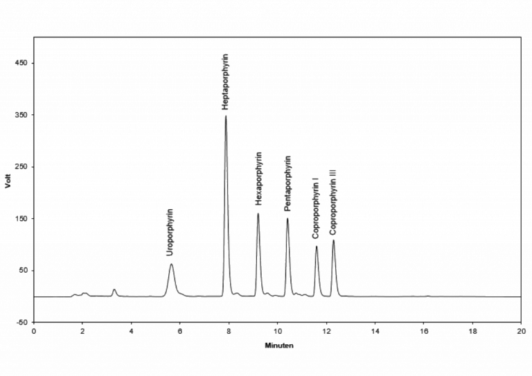
An increase of porphyrins caused by a defect in the synthesis is described as porphyria. The different porphyries are diagnosed by the characteristic pattern of porphyrins in urine. In autosomal dominant acute porphyrin disorders uro-, copro-, penta- und tricarboxyporphyrin are increased. In chronic porphyries including porphyria cutanea tarda the concentration of uro- and heptaporphyrin are increased. In chronic lead intoxication the coproporhyrins are slightly increased, whereas an acute lead intoxication shows extremely high excretion of porphyrins in urine.
Technical data
Sample Urine
Sample volume 1 ml
Detector Fluorescence Ex. 400 nm Em. 620 nm
Method gradient
Determinations 100
Ordering Information
IC2601 Testkit
IC2601ko Controls (2 level each 250 µl lyoph.)
IC2601rp HPLC column
Principle of the method
For the determination of the porphyrins a very easy sample preparation is performed first. The pH value of the sample, calibrator and control is adjusted below 2.5 by addition of some drops (approx. 20 µl) of a stabilization solution. After centrifugation the supernatant is injected into the HPLC system.
The separation via HPLC follows an gradient method at 30 °C, using a „reversed phase“ column; one run lasts 25 minutes. The quantification is performed with the delivered calibrator; the concentration is calculated via integration of the peak areas or peak heights.